Atherosclerosis is one of the most common diseases in the world, it is characterized by a progressive course and leads to severe complications. Characteristic, as a rule, is the involvement of entire arterial system: one patient can be detected atherosclerotic changes in several vascular basins: coronary (cardiac), carotid (arteries of the brain), arteries of the extremities, arteries of internal organs (kidneys, intestines, etc.).The frequency of detection of clinically significant multifocal atherosclerosis is 30-70 % (depending on considered degree of damage). And the difference in clinical manifestations is usually due only to different rate of the disease in different areas. That`s why the patients , who have applied to hospital with a particular arterial pathology, certainly need to assess the condition of other major basins. If necessary, the examination is complemented by contrast angiography(computer or conventional). When a clinically significant lesion of several basins is detected ,the issue of the need for their stage correction , including – surgical correction is discussed.
Sometimes, when seeking medical treatment , the patients don`t even suspect , what can hide behind complaints , and only with a full examination of the patient it is possible to get oriented, to put the right diagnosis and conduct successful treatment.
A patient K.O., 59 y/o, applied to MC Erebouni with complaints of foot coldness, impossibility of walking more than 50 m because of pain in the left leg going away after rest, periodic dizziness. On examination it turned out , that there was no pulse at the level of the inguinal fold on the left, which is a sign of closure of the iliac artery. There was also detected the difference in pressure on the upper extremities of 40 mmHg, what could be evidence of problems with subclavian artery on the left – the main vessel of the hand. The patient was referred to a duplex scan of both lower limbs and upper limbs , as well as the vessels of the neck.
After additional examinations it was detected a gross lesion of the abdominal aorta(constriction 60-65%) and both iliac arteries with occlusion on the left and stenosis on the right to 70 %, stenosis of both femoral arteries 60-70%( in a complex this condition is called Leriche syndrome ). It was detected the stenosis of the left internal carotid artery about 70 %, and also occlusion of the initial segment of the left subclavian artery. In the vertebral artery that is branching from the left subclavian artery was detected retrograde blood flow from the brain to the left arm it is so-called “Seizure syndrome“.
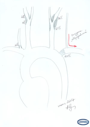 |
(Fig. 1 – A schematic of brachiocephalic artery lesion: stenosis of the right VA 40%, the right ICA 40%, the left ICA 65-70%, occlusion of the first segment of subclavian artery with Seizure syndrome along the left vertebral artery).
Thus, a serious lesion was detected not only in aortoiliac femoral segment, but also in the blood circulation of the brain. Based on the patient`s condition , the Head of the Vascular and Laser Surgery Department Dr. A.S. Badalyan, PhD decided to carry out two interventions on brachiocephalic basin step-by-step. In 23.05.2017 carotid endarterectomy of the left carotid artery was performed with simultaneous carotid -subclavian shunting. The surgery was entirely carried out under the protection of internal shunt at all stages of clamping the vessels to prevent stroke using its own developed tactics.
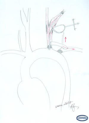 |
( Fig. 2- Brain protection technique. Internal shunt LeMaitre Pruitt-Innahara is installed. The proximal end at the beginning of the procedure was placed below the prosthetic anastomosis. After the procedure of carotid -subclavian shunting it was moved higher with the restoration of blood flow through the subclavian artery).
After 2 weeks the second surgery step was performed: Aorto-femoral bifurcation shunting.
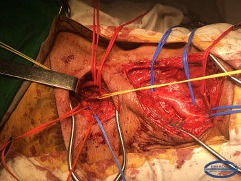 |
Photo 3 Surgical field at the beginning of intervention. Involved vessels are separated and taken on the holders.
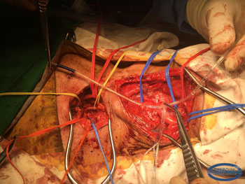 |
Photo 4 The internal shunt before insertion into the artery, is shown in the projection of the artery-a proximal balloon will be located below anastomosis
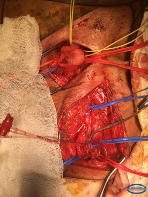 |
Photo 5 The carotid artery is dissected, the internal shunt is placed, and the region is prepared for carotid- subclavian shunting.
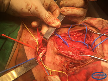 |
Photo 6 The final view of wound
Then in 14.06.2017 the second step of the surgery was performed: Aorto-femoral bifurcation shunting.
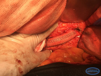 |
Photo 7 Aortic (upper) end of aorto-femoral prosthesis
 |
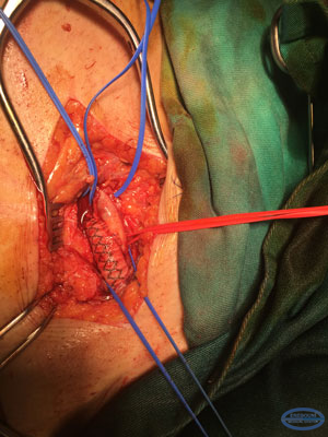 |
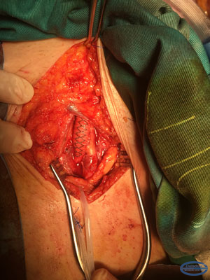
Photo 8,9 Femoral (inferior)ends of aorto-femoral prosthesis.
Both interventions passed without serious complications , an examination performed after the procedure confirmed a full hemodynamic result. The difference in arterial pressure on the hands after the surgery is acceptable (5-10 mm Hg), the patient is able to walk more than 500 meters.

