Despite of significant achievements in the sphere of diagnosis and treatment of acute surgical diseases, in recent decades, there are poor prognoses yet for the patients with acute thrombosis of mesenteric artery that frequently (in 59-93% cases) are accompanied by in-hospital mortality. In more than 20% of cases of thromboembolism of mesenteric arteries, emboli are multiple, located in various arteries and organs, which even worsens the prognosis of the disease.
If there is a suspicion of thromboembolism of the artery of one or another organ of the abdominal cavity, first of all it is necessary to exclude mesenteric artery embolism. Rapid diagnosis, correct surgical approach and intensive treatment can significantly improve prognosis and reduce mortality.
In 06.03.17.,a patient A.E.,72 y/o has admitted to Emergency Medical Care Department in severe condition and was hospitalized to Intensive Care Unit.
Complaints appeared and developed a day before coming to the hospital (according to the anamnesis of the disease patient has atrial fibrillation and post-thrombophlebitic syndrome).
CT angiography revealed a picture of stenosis of the abdominal aorta and its branches, embolism of the right branch of the superior mesenteric artery (Fig. 1), embolism of the right common femoral artery (Fig. 2), post infarction cystic formations in the spleen.
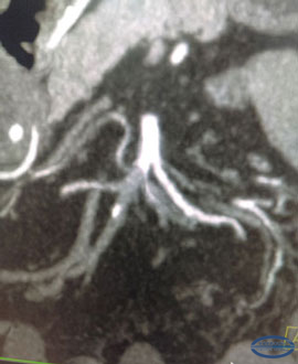 |
Fig. 1.
CT images of the embolism of the right branch of the superior mesenteric artery
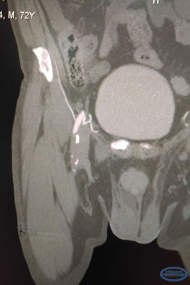 |
Fig.2.
CT images of post infarction cystic formations in the spleen.
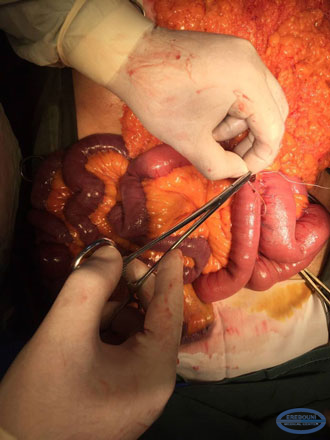
In 06.03.17 due to strict life-threatening indications, the median laparotomy was carried out by the team of general surgeons of the General & Thoracic Surgery Department headed by Dr.Vardanyan A.S., PhD (c.m.s.). During intraoperative inspection the necrosis of the distal 2/3 of the jejunum, the entire ileal and right half of the colon was revealed (Fig. 3).
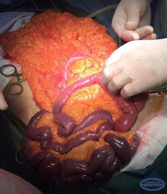 |
Fig. 3.
Intraoperative inspection showed necrosis of the distal 2/3 of the jejunum, entire ileal and right half of the colon
Viable was only the proximal part of the jejunum for a long of 50-60 cm from the Treitz` ligament, and also in the left half of the large intestine, including the sigmoid colon and rectum (Fig. 4).
|
|
Fig. 4.
The viable part of the intestine
The resection of necrotic intestinal parts with the formation of jejuno-transverse anastomosis side-by-side with 2-row nodal sutures was performed.
After closure of laparotomy wound, a team of vascular surgeons under the Head of Vascular and Laser Surgery Department Dr. A. Badalyan MD, Ph.D, endarterectomy of the right common and deep femoral artery, profundoplasty with an alloy prosthesis was performed (chronic occlusion of the superficial femoral artery from its ostium was revealed, the common femoral artery was also obstructed by embolus; there was revealed the significant fibrocalcinosis of blood vessels, including deep femoral artery up to 5 cm).(Fig. 5).
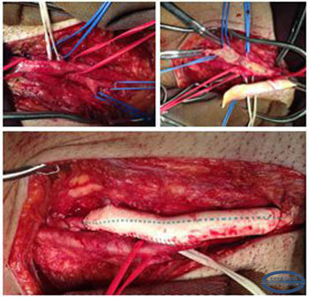 |
Fig. 5.
Endarterectomy of the right common and deep femoral artery, profundoplasty with alloy prosthesis
The postoperative period was uneventful, intestinal activity was restored on the 3rd day. Acute ischemia of the right lower limb was completely solved without postischemic complications. After repeated duplex scanning positive results, the patient was transferred to the general surgery department, and was discharged on the 10th day with recovery.
Acute hypoperfusion of the intestine can be of 4 types. The most common cause (40-50% of cases) is arterial embolism. Cardiogenic embolism is the most common cause of acute mesenteric ischemia. By frequency of occurrence, the second place is an acute mesenteric thromboses (25-30%). Non-occlusive mesenteric ischemia occurs in a low cardiac output combined with diffuse mesenteric vasoconstriction. Thrombosis of mesenteric veins is a relatively rare cause of ischemia (just over 10%) of the abdominal organs., The superior mesenteric artery is most vulnerable to embolism due to the acute angle with the aorta and its large diameter.
The conducted researches revealed only 2 cases when combined embolization of the mesenteric artery and lower extremity with acute ischemia were described (in the literature the case of combined thromboembolism of the superior mesenteric artery and brachial artery was described, when late diagnosis led to amputation of the upper limb).
Highly complex and unique surgical intervention, carried out by a multidisciplinary team of highly qualified surgeons of the Erebouni MC, with the correct definition of tactics and sequence of the multiprofile intervention, not only saved the patient's life, but also prevented limb amputation and his further disability!

