Urolithiasis disease (kidney stone disease) today is one of the most actual and frequent problems of urology. The most severe form of kidney stone disease is the kidney staghorn calculi that occurs in 5 to 25% cases.
The main surgical interventions in urolithiasis that performed in the Department of Urology of MC Erebouni include:
- Remote lithotripsy
- Endoscopic destruction and removal of stones from the renal pyelocaliceal system and the urinary system on any levels
- Endoscopic destruction of bladder stones
- Percutaneous nephrolithotripsy (PNL)
- Retrograde intrarenal surgery with the use of laser lithotripsy
The peculiarity of the staghorn nephrolithiasis is that it fills all cavities of pyelocaliceal system (PCS) and therefore has the form of coral. Staghorn calculi impede the outflow of urine, but not for a long time, that prevents the formation of new and smaller concrements.
As a result, the staghorn nephrolithiasis is a severe pathology, insidious with its complications and requiring an immediate treatment.
Gradually, with the increase of the size of the stone, the patient starts complaining to dull aching pain in the lower back. But these symptoms occur so imperceptibly that people hardly can determine the time of their occurrence.
Today in the world the percutanious nephrolithotripsy has firmly established itself as a standard method of treatment of this category of patients, providing clinical efficacy in 88-98% of cases.
In the Department of Urology of Erebouni MC we do not perform, open surgeries in staghorn nephrolithiasis.
At present percutanious nephrolithotripsy is recognized as a leading method of surgical treatment of staghorn nephrolithiasis and it makes 99.9% of all surgical interventions in this pathology. In the Department of Urology of MC Erebouni this intervention has been applied since 2006.
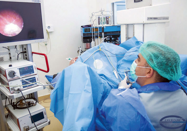
One of the main conditions for carrying out this category of surgeries - is an appropriate contemporary technical facilities and medical equipment in the operating room including the X-ray endoscopic set, ultrasonic diagnostic equipment, endoscopic equipment, personnel radiation protection system, appropriate supply of medicines required for the rehabilitation of patients after intervention.
In Mc Erebouni X-ray endoscopic room is equipped with modern X-ray set of Siemens Access Uroskop, Siemens ultrasound diagnostic equipment, Richard Wolf endoscopic stand (Germany), Swiss LITHOKLAST and LITHOKLAST-MASTER (EMS Swiss) for kidney stones contact breaking, Richard Wolf laser Megapulse (Germany), Carl Stotz endoscopic instruments (Germany), allowing to remove stones from any levels of the urinary system.
The classic surgery is carried in the lying on the stomach position of the patient. In the Department of Urology of Erebouni MC has already performed more than 1000 such kind of surgeries.
Patient`s body weight has been from 55 to 158 kg. In 90% of cases spinal and epidural anesthesia is applied, which is carried out by a qualified doctor- anesthesiologist, it is well- tolerated by the patients, allows prolonging the analgesic effect in postoperative period.
With the help of ultrasound guidance the puncture of renal pyelocaliceal system is performed, which significantly minimizes radiation exposure of a patient and a surgeon. After the puncture of renal pyelocaliceal system, the guidewire is delivered through a needle tube, on which the further formation of nephrostomy channel is carried out.
With the help of a plastic bougies the dilatation of the wound chanal is implemented and Amplatz sheath is placed. Through this sheath the probe lithotripter is introduced and removing of stone is performed.
With the help of a combined lithotripter, a breaking up the kidney stones and simultaneous its evacuation are carried out. After breaking up and removing the stone fragments the nephrostom tube is set into renal pyelocaliceal (collecting) system of the kidney for adequate urinary diversion.
This type of surgery allows removing the stones of different sizes from renal pyelocaliceal system. It is minimal traumatic and well- tolerated by the patients. The rehabilitation period is about 1 to 5 days.
Postoperative period requires gentle nutrition with low content of calcium, phosphorus, and various condiments. The treated patients stay under the follow up control of the specialists of the department who give recommendation and workout the individual for each patient diet. Regular preventive examination of the kidney provides healthy life for a long time.


