
In MC Erebouni within the framework of the two-day conference (9-10 June, 2017) master classes by prominent endoscopists from St Petersburg were held. Many specialists in the field of endoscopy, thoracic and general surgery were invited.
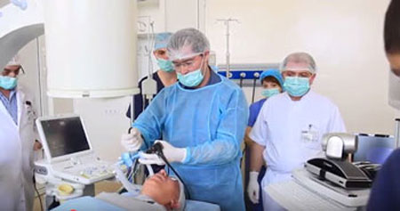
On June 9, 2017 three patients with various pathologies of gastrointestinal tract were carried out the rarest surgical interventions with the help of last generation endoscope of expert class and EUS (endoscopic ultrasound), that was brought and presented by Russian branch of PENTAX company. The Head of endoscopy department of Research Institute of Surgery and Emergency Medicine, Pavlov State Medical University of St. Petersburg, Dr. Smirnov A.A., (PhD) conducted master-class with demonstration of diagnostic, surgical and endoscopic methods in this field. It is important to note, that in Russia such surgeries are carried out only by two specialists, one of them is Dr. Smirnov A.A.. Cameras mounted in a conference hall and operating room allowed to monitor the surgery online, which were interactively highlighted by Dr. Smirnov A.A.
Certainly, such kind of interventions should be carried out using modern technical equipment, with a high professional anesthesiology service and careful post-operative follow-up of patients. It is extremely important to note the fact, that such kind of surgeries have higher rate of rapid rehabilitation and early return to the usual rhythm of life, without affecting its quality.
Endoscopic examinations are carried out with the help of digital video-endoscopic system of the last generation- PENTAX Hi-Line HD +(Japan). Similar master -class was held earlier in October 2016 in MC Nairi (it was also organized by Russian branch of PENTAX company).
Endoscopy - is the method of examination of internal organs with the help of a special instrument (endoscope). Gastrointestinal endoscopy is one of the leading and best methods of diagnosis without surgical intervention, and the most frequent method of instrumental examination of the patients. The examination is carried out by an endoscope, with an eyepiece at one end, that allows to visualize an image of the examined organ, and the camera - at the other end, transmitting the image from various gastrointestinal areas. Experienced specialists can detect a problem at an early stage of development and timely prescribe a necessary treatment. At present there are no contraindications for endoscopy. Modern technologies allow to perform stomach endoscopy under general anesthesia. Such procedure is carried out under control of anesthesiologist. During and after the procedure the patient doesn`t feel discomfort or any unpleasant feelings. The examinations are short, however, it should be taken into consideration that the duration of each procedure is individual and depends on a number of factors.
(In more details it was highlighted in an article at the follow the link https://www.erebunimed.com/rus/news.more/197)
Performing ultrasound examination from the inside, being in close proximity to the examining organs - EUS allows to examine internal organs in details, by seeing the changes that are not available for standard ultrasound examination. Examinations are carried out with the help of a special endoscope, at the end of which is not only an optic instrument, but also a tiny ultrasonic sensor. EUS allows to perform ultrasound examination of the hollow organs of the GI tract, in which echoendoscope is inserted (walls of esophagus, stomach, colon and rectum), as well as abdominal organs, which are located nearby to stomach or intestine (pancreas, bile ducts, gallbladder and liver).
Ultrasound scan through esophagus makes possible to receive an image of mediastinal organs as well as to determine the condition of lymph nodes situated in abdominal or chest cavity. Sonoelastography during EUS allows to characterize and differentiate benign and malignant changes of lymph nodes with a high sensitivity, specificity and accuracy. Sonoelastography image complements the other modes of scanning in EUS and allows to carry out fine needle aspiration biopsy in case of multiple lymph nodes involvement.
The first patient: Ds - tumor of pancreas. EUS alongside with puncture biopsy were performed.
The second patient: achalasia of esophagus in the compensation stage. Esophagogastroduodenoscopy was performed (erosion was determined: tissue sample was taken for biopsy).
The third patient was examined by the same method for the formation in the stomach (the patient was offered dynamic examination).
( About indications for performing and diseases in which the above diagnostic methods are applied : follow the link https://www.erebunimed.com/rus/news.more/197)
Endoscopic ultrasound favorably differs from all the above methods, allowing at an early stage to detect a number of serious diseases, in order to increase the effectiveness of treatment. The device for endoscopic ultrasound allows to perform ultrasound examination of internal organs, including areas, that are inaccessible for a standard ultrasound. Unlike computed tomography, endoscopic ultrasound makes it possible to examine internal cavities in various planes, following the direction of the duct. Besides, when performing EUS, visually-controlled (with the help of ultrasound) targeted biopsy of a certain site could be carried out. All these advantages of diagnostic and surgical endoscopy over the other methods raise diagnosis and treatment to a qualitatively new level.
The second day of the conference - June 10, 2017 was dedicated to bronchoscopic examinations. Topic: “Modern possibilities of bronchoscopy in diagnoses of pathology of lungs and mediastinum”. To participate in the conference was invited thoracic surgeon, the head of intervention pulmonology Department of St. Petersburg State Research Institute of Phthisiopulmonology Igor. V. Vasilyev, MD, PhD. Together with him the thoracic surgeon of MC Erebouni Dr. G.B. Melikyan, who has recently undergone further training in this field exactly in that scientific institution, took part in the surgery.
With a salutatory word, Executive Director of MC Erebouni M.B. Manukyan, as well as Head of General & Thoracic Surgery Clinic, Dr. Hovhannisyan A.K. MD., PhD, Grand PhD (d.m.s.) addressed to the audience. They noted the importance and necessity of implementation and usage of modern medical achievements in our republic. They expressed their pride in the fact, that such innovations for the first time in Armenia are implementing in MC Erebouni.
Several patients with various pathologies were performed video-diagnostic bronchoscopy with different I-scan modes.
The first patient (with suspected central lung cancer). The purpose: demonstration of the technique of diagnostic video-bronchoscopy and I-scan functions.
The second patient was carried out a procedure: “EBUS-TBNA (endobronchial ultrasound – transbronchial needle aspiration) to a patient with mediastinal lymphadenopathy”. The purpose: demonstration of the technique of examination of lymph nodes and biopsy of 7 groups, technique of medicine preparation.
The third patient was carried out a procedure: “EBUS-TBNA in patient with lung cancer”. The purpose: demonstration of the technique of systemic staging by using EBUS-TBNA”.
The fourth patient: with peripheral formation of superior lobe of the left lung. He was carried out video-bronchoscopy and transbronchial lung biopsy.
As on the first day of the conference, on the second day all surgeries in online mode were broadcast directly from operating room.
It is very important to note, that the EBUS-TBNA procedure was first performed in Armenia, in MC Erebouni.
Widespread implementation of the above method into the practice, morphological verification of hard-to-reach tumors of lungs and mediastinum, as well as their staging will help to diagnose these diseases at a more qualitative level, and therefore to choose the right tactics and treatment strategy.